Category: Uncategorized

  • Wireless Skin Analyzer Dermatoscope: A Practical Guide to Digital Skin Magnification and UV Inspection

    Wireless Skin Analyzer Dermatoscope: A Practical Guide to Digital Skin Magnification and UV Inspection

    Skin is a complex surface that can show differences in texture, pigmentation, hair growth, and other visible characteristics. For skincare professionals, beauty practitioners, educators, and people interested in skin observation, magnification can provide a closer look at details that are difficult to see under ordinary lighting. Digital inspection tools make this process more accessible by combining a magnifying optical system with electronic imaging and, in some cases, wireless connectivity.

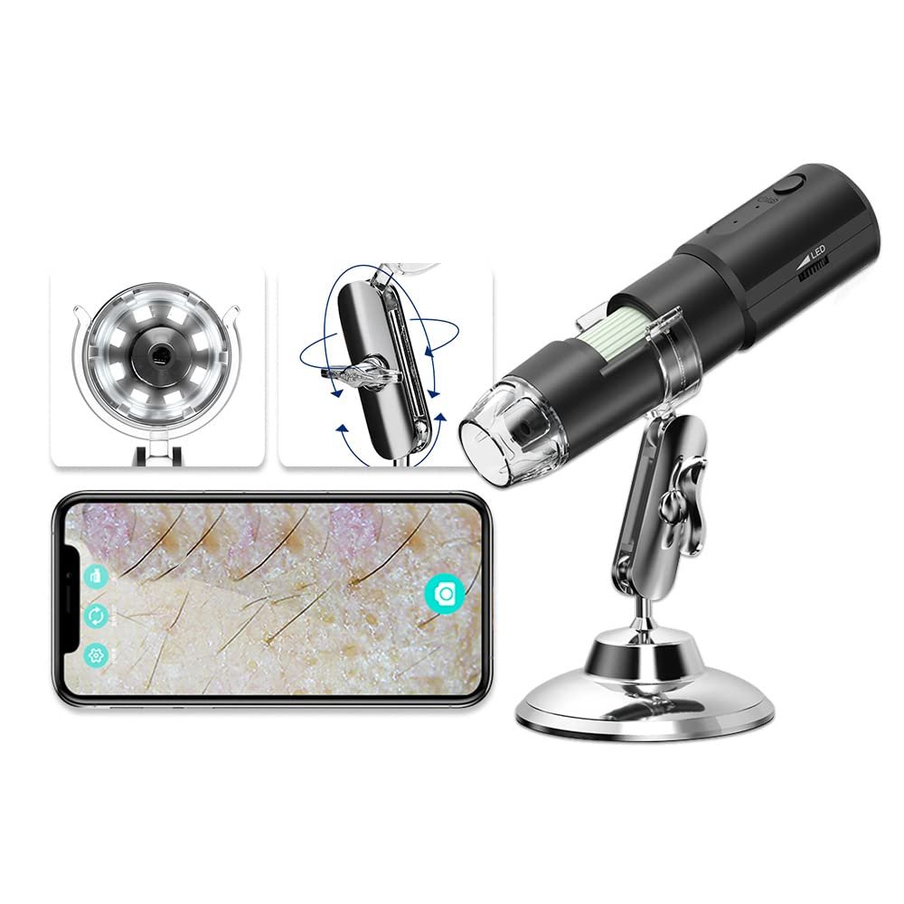
    The Wireless Skin Analyzer Dermatoscope UV 1000X Magnifier Digital Microscope is designed around this general concept. Its product title highlights digital magnification, wireless operation, UV-related illumination, and a dermatoscope-style inspection approach. These features place it within a category of devices intended to display enlarged images of skin or other surfaces for closer visual examination.

    Tools of this kind can be relevant to skincare education, cosmetic observation, skin-surface documentation, and certain professional workflows. However, it is important to understand the distinction between viewing skin at high magnification and medically evaluating a skin condition. An enlarged image alone cannot establish a diagnosis, determine whether a lesion is cancerous, or replace an examination by a qualified healthcare professional.

    This guide explains the main concepts behind digital skin analyzers, how magnification and illumination can be used, what wireless connectivity may contribute, and the practical considerations that matter when choosing or using a skin inspection device.

    What Is a Digital Skin Analyzer and Dermatoscope?

    A digital skin analyzer is an imaging device designed to magnify a surface and display the resulting image electronically. Depending on the model, it may use a camera sensor, integrated illumination, a digital display or connected smartphone, and software for viewing or saving images.

    A dermatoscope is a related but more specialized instrument used in clinical skin examination. Traditional dermatoscopes commonly use magnification and controlled illumination to help trained healthcare professionals examine skin structures that are not easily visible to the unaided eye.

    Although the terms can overlap in product descriptions, not every digital skin analyzer is equivalent to a clinical dermatoscope. Medical dermatoscopy depends on suitable optics, illumination, image quality, and professional interpretation. A consumer-oriented digital microscope may provide enlarged pictures without offering all the capabilities required for clinical assessment.

    The Wireless Skin Analyzer Dermatoscope UV 1000X Magnifier Digital Microscope is described as a wireless digital magnification tool with UV-related functionality. Its exact optical specifications, supported devices, illumination wavelength, and intended professional applications should be confirmed in the current product documentation.

    Understanding those details is particularly important when a device is marketed for skin inspection, because the intended use determines which observations it can reasonably support.

    Key Features of a Wireless Skin Analyzer

    Digital Magnification

    Magnification is one of the main reasons people use digital microscopes and skin inspection tools.

    An enlarged image can make surface features easier to observe, including texture, pores, hair shafts, and visible pigmentation patterns. This may help users compare different areas of skin or document changes in appearance over time.

    The product title references magnification up to 1000X. That figure should be interpreted carefully because advertised magnification can depend on optical magnification, digital enlargement, display size, and the method used to calculate the figure.

    Digital enlargement can make an image appear much larger without necessarily revealing additional detail. Actual resolution depends on the camera sensor, lens quality, focus, illumination, and image processing.

    For practical inspection, a clear and well-focused image is generally more useful than a large but blurry image.

    Wireless Connectivity

    Wireless operation can make a digital inspection device more flexible.

    Depending on the product’s design, wireless connectivity may allow an image to be viewed on a compatible smartphone, tablet, or other supported device. A larger display can be useful when examining fine details or discussing an image with another person.

    Wireless imaging may also help users capture and organize observations if the device and its software support image storage.

    However, wireless connectivity should not be assumed to include every possible feature. Live viewing, photography, video recording, file transfer, and compatibility with different operating systems are separate capabilities that should be checked in the product documentation.

    Before relying on a particular workflow, confirm whether the device supports the intended phone or tablet and whether it requires a dedicated application.

    UV-Related Illumination

    The product title identifies UV functionality, which makes illumination safety an important consideration.

    Ultraviolet radiation is a form of electromagnetic radiation that is not fully visible to the human eye. UV sources vary considerably in wavelength and intensity, and different wavelengths can have different effects on skin and eyes.

    A product described as having UV illumination may use a particular UV source for surface observation, but the exact wavelength, intensity, exposure guidance, and intended purpose should not be inferred from the product title alone.

    Users should consult the manufacturer’s specifications to understand what type of light the device produces and how it is meant to be used.

    UV illumination should not automatically be interpreted as a diagnostic feature. It does not establish that a skin condition is harmless or serious, and it should not be treated as a substitute for clinical examination.

    Digital Image Viewing

    A digital skin microscope can provide an enlarged image on a connected display rather than requiring the user to look through a conventional optical eyepiece.

    This format can make observations easier to share and document when the necessary software features are available.

    For skincare education, for example, a practitioner may want to show the appearance of a skin surface while explaining general concepts such as texture, dryness, or visible pores.

    The image should still be interpreted within the limits of the device. Lighting, camera angle, magnification, and image processing can influence how the surface appears.

    Portable Inspection Format

    Wireless digital microscopes are often designed to provide a compact alternative to larger laboratory imaging equipment.

    A portable device can be convenient for demonstrations, personal observation, training, and other tasks where a full-size microscope would be unnecessary.

    The actual portability of this particular model depends on its dimensions, power source, battery arrangement, and included accessories. Those details should be verified before planning to use it away from a desk or dedicated workspace.

    How Digital Skin Magnification Works

    Understanding the basic imaging process can help users interpret what they see.

    A digital microscope directs light toward a surface and uses an optical system to capture the reflected light. A camera sensor converts that information into an electronic image, which is then processed and displayed.

    The final image is influenced by several factors.

    Optics: The lens determines how light is collected and focused.

    Sensor: The camera sensor affects how much image detail can be captured.

    Illumination: Light intensity, angle, and wavelength can change the visibility of surface features.

    Focus: The lens must be positioned correctly to produce a clear image.

    Digital processing: Software may adjust contrast, color, sharpness, or image size.

    These factors work together. A device advertised with high magnification may still produce limited detail if its sensor or optics cannot resolve very small structures.

    For skin observation, consistent lighting and focus can be especially important because the surface is not perfectly flat. Different areas may also reflect light differently.

    How the Wireless Skin Analyzer Can Be Used

    General Skin-Surface Observation

    A digital skin analyzer can be used to view surface characteristics more closely than is possible with the naked eye.

    Examples include visible texture, dryness, fine lines, pores, and hair shafts.

    These observations may be useful for cosmetic education or general awareness of the skin’s appearance.

    However, a close-up image cannot independently establish the cause of a visible feature. Dryness, redness, pigmentation, and surface irregularities can have many possible explanations.

    People who notice persistent or concerning skin changes should seek appropriate medical advice rather than attempting to diagnose the cause from magnified images.

    Skincare Education

    Skincare educators and beauty professionals may use magnification to explain general concepts related to skin appearance.

    For example, an enlarged image can help illustrate the difference between a smooth-looking surface and one with visible texture or dryness.

    This may make a demonstration more interactive because the user can see the feature being discussed rather than relying entirely on a verbal description.

    The tool should be used for education within the practitioner’s qualifications and should not be presented as proof of a medical diagnosis.

    Cosmetic Consultation Support

    In a cosmetic setting, an imaging device may help a practitioner document visible surface characteristics or discuss a client’s general skincare concerns.

    If images are captured, consistent lighting and positioning can make comparisons more meaningful.

    However, image appearance can change with camera distance, focus, illumination, skin moisture, and image processing. A difference between two images does not necessarily mean that a product or treatment caused a change.

    Any claims about treatment effectiveness should be based on appropriate evidence rather than an informal comparison of magnified images.

    Comparing Skin Appearance Over Time

    Some users may be interested in documenting the appearance of a particular area over time.

    If the device supports image capture and storage, it may provide a way to keep visual records.

    For a meaningful comparison, images should be captured under similar conditions. Differences in lighting, magnification, angle, and focus can create misleading impressions.

    Skin documentation should also be handled with care because images of the body can be sensitive personal information.

    For changes involving moles, lesions, unusual pigmentation, or other potentially concerning features, photography should not delay a medical appointment.

    Educational Demonstrations of Hair and Skin Structure

    Magnification can be useful when explaining general surface structures, including the appearance of individual hair shafts or skin texture.

    The quality of the observation depends on the microscope’s actual resolution and optical design.

    Users should avoid interpreting every visible variation as a medical problem. Normal skin differs between people and across different body areas.

    General Surface Inspection Beyond Skin

    Some digital microscopes can also be used to inspect non-biological surfaces, such as fabric, paper, small manufactured components, or other objects that fit within the device’s working distance.

    Whether this is appropriate for the particular model depends on its intended use, optical arrangement, and the manufacturer’s guidance.

    If the device is used on non-skin objects, users should follow any cleaning instructions before returning it to skin-related use.

    Who May Find This Type of Device Useful?

    Skincare Educators

    Educators who explain general skin-surface concepts may find digital magnification useful for demonstrations.

    A displayed image can help illustrate texture and other visible characteristics, provided that the discussion remains within the limits of the tool.

    Beauty and Cosmetic Practitioners

    Professionals working in cosmetic settings may use suitable imaging tools to support visual demonstrations or document appearance.

    They should confirm that the device is appropriate for their intended workflow and avoid making medical claims beyond their qualifications.

    Students Learning About Skin

    Students in relevant educational programs may benefit from seeing enlarged images of surface features.

    A digital microscope can provide a visual introduction to magnification, lighting, and basic imaging principles. It should not be treated as a substitute for formal clinical training or specialized medical equipment.

    People Interested in Personal Observation

    Some individuals may want to learn more about the visible characteristics of their skin.

    A digital magnifier can support general observation, but it is important to avoid interpreting enlarged images as diagnostic results.

    If the purpose is to evaluate a medical concern, a qualified healthcare professional is the appropriate source of assessment.

    Users Who Need Digital Image Sharing

    Wireless imaging may be useful when the device supports viewing on a compatible screen or sharing captured images.

    This can support demonstrations, education, or documentation. Users should consider privacy and consent before capturing or sharing images of another person’s skin.

    Important Things to Consider Before Using a Skin Analyzer

    A Magnified Image Is Not a Diagnosis

    This is the most important limitation to understand.

    A skin analyzer can show enlarged visual information, but it cannot necessarily determine whether a mole, lesion, rash, or other skin change is benign or medically concerning.

    Clinical assessment can involve medical history, examination, dermatoscopic interpretation, and additional testing when appropriate.

    A device advertised with 1000X magnification should not be assumed to provide diagnostic accuracy simply because it produces a highly enlarged image.

    If a spot is changing in size, shape, or color, or if a lesion is bleeding, persistently itching, painful, or otherwise concerning, seek medical advice. Do not rely on a consumer microscope to rule out a serious condition.

    Verify the Actual Magnification and Resolution

    Advertised magnification figures can be difficult to compare across digital devices.

    Some figures may include digital enlargement, which increases displayed image size without necessarily increasing the amount of detail captured.

    Look for reliable information about the sensor resolution, optical design, focus range, and image quality.

    The practical question is not only how large an image can become, but whether important details remain clear and distinguishable.

    Understand the UV Specifications

    UV-related features require particular attention.

    Confirm the type of UV source, its wavelength, intended use, and any exposure precautions described by the manufacturer.

    Do not look directly into a UV source or expose skin or eyes to UV radiation in a way that is inconsistent with the product instructions.

    If the device does not provide clear safety information, seek clarification from the manufacturer before using its UV function.

    The presence of UV illumination does not establish that a device can detect skin cancer, diagnose infection, or reliably identify a particular skin condition.

    Skin Sensitivity and Hygiene

    Any device that comes into contact with skin should be handled according to the manufacturer’s hygiene instructions.

    Clean the device as directed, and avoid applying unapproved chemicals to optical components or surfaces that touch the skin.

    If a device is used by multiple people, confirm whether the manufacturer permits that arrangement and follow appropriate cleaning and disinfection procedures.

    Do not use the device on broken, irritated, or infected skin unless a qualified healthcare professional has advised that the intended use is appropriate.

    Wireless Compatibility

    Wireless functionality can vary between devices.

    Check whether the microscope supports the operating system, phone, tablet, or computer that you intend to use.

    Also confirm whether the required application is available, whether an account is necessary, and whether image storage or sharing requires additional software.

    Wireless connection alone does not guarantee that every image can be saved or transferred in the desired format.

    Privacy and Image Storage

    Skin images can contain sensitive personal information.

    If images are stored on a smartphone, tablet, or cloud service, consider who can access them and how long they will be retained.

    Before capturing or sharing another person’s skin images, obtain appropriate consent.

    Professionals should follow applicable privacy requirements and workplace policies when documenting clients or patients.

    Lighting and Image Interpretation

    Illumination can change how the skin appears.

    Direct light may emphasize texture, create glare, or make certain areas look more prominent. Different camera angles can also alter the appearance of surface features.

    When comparing images, try to maintain consistent positioning and lighting.

    Even under controlled conditions, visual differences should not automatically be attributed to a particular product, treatment, or health condition.

    Power and Maintenance

    Digital imaging devices need an appropriate power source and careful handling.

    Check the manufacturer’s instructions for charging, battery use, cleaning, storage, and operating conditions.

    Protect the lens from scratches and dust. Avoid forcing adjustment mechanisms or exposing electronic components to moisture unless the device is specifically designed for such conditions.

    Digital Skin Analyzer vs. Traditional Magnifying Glass

    A traditional magnifying glass is simple, portable, and generally does not require power.

    It can be useful for quickly examining surface details or reading small text.

    A digital skin analyzer provides a different experience by presenting an enlarged image on a screen, potentially with integrated lighting and image-capture capabilities.

    This can be useful when a user wants to inspect an area while viewing the image on a separate display.

    However, a magnifying glass may be easier for quick, informal observations, while a digital microscope offers electronic imaging features that may be helpful for documentation.

    Neither tool independently provides a medical diagnosis.

    Digital Skin Analyzer vs. Clinical Dermatoscope

    A clinical dermatoscope is designed for trained healthcare professionals to examine skin structures under controlled optical conditions.

    Depending on the model, it may include specialized illumination, optical magnification, and features intended for clinical examination.

    A digital skin analyzer may focus more on displaying enlarged images through a camera and screen.

    These categories can overlap, but they should not be treated as equivalent without confirming the product’s specifications and intended use.

    Anyone seeking a tool for clinical dermatology should consult qualified professionals and select equipment appropriate for the specific medical application.

    Digital Microscope vs. Smartphone Camera

    A smartphone camera is convenient because it is already available to many users.

    It can capture ordinary photographs and, in some cases, provide macro photography or digital zoom.

    A dedicated digital microscope is designed specifically for close-up viewing and may provide a more controlled working distance, integrated illumination, and specialized optics.

    The difference depends on the particular equipment. Some smartphones have capable macro systems, while some compact microscopes have limited resolution.

    The appropriate option depends on the detail required, the intended workflow, and whether a dedicated inspection device provides meaningful advantages.

    How to Get More Consistent Images

    Use Stable Positioning

    Movement can blur close-up images. Place the device on a stable surface or use its intended support system.

    If the microscope is handheld, keep the device and subject as steady as possible.

    Adjust Focus Carefully

    Small changes in distance can affect sharpness significantly at high magnification.

    Follow the device instructions for focusing rather than relying on digital enlargement to compensate for a blurred image.

    Control the Lighting

    Avoid excessive glare and harsh reflections.

    If the image appears washed out, adjust the illumination or the angle of the subject where possible.

    Keep Comparisons Consistent

    If the goal is to compare images, maintain similar lighting, camera distance, angle, and focus.

    This makes the images easier to interpret and reduces the likelihood of mistaking a photographic difference for a real change in the skin.

    Avoid Overinterpreting Small Details

    Magnification can reveal normal surface variations that are difficult to see without assistance.

    Not every irregularity is a medical problem, and not every apparently normal image excludes one.

    Use the device for observation rather than self-diagnosis.

    Frequently Asked Questions

    What is a wireless skin analyzer dermatoscope?

    It is a type of digital inspection device intended to magnify skin or surface details and display the resulting image electronically. Wireless models may connect to compatible devices for viewing or other supported functions.

    What does 1000X magnification mean?

    The product title advertises magnification up to 1000X. The practical meaning depends on how magnification is calculated, including whether digital enlargement is involved. The number alone does not establish the amount of fine detail the device can resolve.

    Can this device diagnose skin cancer?

    A consumer digital skin analyzer should not be assumed to diagnose skin cancer. Diagnosis requires appropriate clinical assessment by a qualified healthcare professional. Do not use magnified images to rule out a concerning lesion.

    What is the purpose of UV illumination?

    UV illumination can be used for particular surface-observation applications, depending on the source wavelength and device design. Its exact purpose and safety requirements should be confirmed in the manufacturer’s documentation.

    Is UV illumination safe for skin and eyes?

    Safety depends on the wavelength, intensity, exposure duration, and intended use. Follow the manufacturer’s precautions, avoid looking directly into the UV source, and do not expose skin or eyes beyond the specified instructions.

    Can it connect to a smartphone?

    Wireless connectivity is indicated by the product title, but the supported devices, operating systems, applications, and connection process should be checked in the current product documentation.

    Can images be saved?

    Image-saving capabilities vary by model. Confirm whether the device supports still photographs, video recording, file transfer, or other storage features before relying on it for documentation.

    Can a skin analyzer be used for cosmetic consultations?

    A suitable device may support visual demonstrations or documentation of surface appearance. It should not be used to make medical diagnoses or claims beyond the practitioner’s qualifications.

    Can it replace a dermatologist’s examination?

    No. Digital magnification can provide additional visual information, but it cannot replace a clinical examination when a medical concern needs assessment.

    Does higher magnification always mean a clearer image?

    No. Image clarity also depends on resolution, optics, lighting, focus, and digital processing. High enlargement can make an image bigger without revealing more useful detail.

    How should the device be cleaned?

    Follow the manufacturer’s cleaning and disinfection instructions. Use only approved methods, especially for lenses and any surfaces that contact skin.

    Is it suitable for professional medical use?

    That depends on the device’s intended use, specifications, regulatory status where applicable, and the requirements of the clinical application. Professionals should verify those details rather than relying on the product title alone.

    Conclusion

    The Wireless Skin Analyzer Dermatoscope UV 1000X Magnifier Digital Microscope belongs to a category of digital inspection tools designed to make small surface details easier to observe. Its listed features point toward magnification, wireless imaging, and UV-related illumination, which may be relevant to certain skincare education, cosmetic observation, and general inspection workflows.

    The practical value of such a device depends on its actual optical quality, sensor resolution, focus range, illumination characteristics, supported connections, and intended use. The advertised magnification figure should not be treated as a guarantee of diagnostic precision.

    UV specifications and safety instructions also deserve careful attention. Users should confirm the type of UV source and follow the manufacturer’s guidance for exposure and operation.

    Most importantly, magnified images are not a substitute for medical assessment. A digital microscope may help users observe visible features, but it cannot reliably determine whether a lesion is harmless or serious without appropriate clinical evaluation.

    For people interested in skin-surface observation or digital magnification, understanding the device’s capabilities and limitations is the best starting point. Confirming the specifications, using the equipment as directed, protecting sensitive images, and seeking professional medical advice when needed can help ensure that the technology is used appropriately.
    You can check more details on Amazon here.

  • Elikliv Microscope: A Practical Guide to the 4.3-Inch Digital Coin Magnifier With Lights

    Elikliv Microscope: A Practical Guide to the 4.3-Inch Digital Coin Magnifier With Lights

    Small objects often contain details that are difficult to see with the naked eye. A coin may have tiny lettering around its edge, a collectible stamp can show subtle printing differences, and an electronic circuit board may contain components that are almost impossible to inspect comfortably without magnification. For collectors, hobbyists, students, and people who work with small objects, a digital microscope can make these details easier to examine.

    Unlike a traditional optical magnifier, a digital microscope uses a camera sensor to capture an enlarged image and presents it on a screen. This changes the way users interact with the object. Rather than holding a small lens close to the eye, they can look at a display while adjusting the object, lighting, and magnification to suit the task.

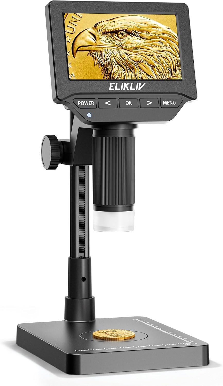
    The Elikliv Microscope, described as a 4.3-inch digital coin magnifier with lights for adults, is designed around this approach. Its screen-based format is intended to help users inspect coins and other small objects while providing illumination for close-up viewing. This type of device can also be relevant to people interested in stamps, jewelry, electronic components, small mechanical parts, and educational observation.

    The usefulness of a digital microscope depends on more than magnification alone. Lighting, focus, working distance, image clarity, stability, screen size, and the characteristics of the object being examined all affect the final result.

    This guide explains how the Elikliv digital microscope fits into the broader category of compact inspection tools, how it may be used for coin collecting and other hobbies, what to consider before using one, and how digital microscopes compare with traditional magnifiers and more advanced optical equipment.

    What Is a Digital Microscope?

    A digital microscope is an optical inspection device that uses a camera sensor and digital display to show enlarged views of small objects.

    A conventional magnifying glass relies on a lens to enlarge the image viewed directly through it. A digital microscope instead captures the image electronically and displays it on a screen. Depending on the device, the image may also be available for photography, recording, or transfer to another device.

    This screen-based arrangement can make extended inspection more comfortable for some users because they do not have to position their eye directly behind a small eyepiece.

    Digital microscopes are available in many forms, ranging from compact handheld units to larger laboratory instruments with adjustable stands, external monitors, and specialized lighting systems.

    A compact model with a 4.3-inch screen is oriented toward personal inspection and everyday hobbies rather than necessarily replacing a professional laboratory microscope.

    For coin collectors, the main appeal is the ability to examine small features such as mint marks, lettering, surface texture, fine scratches, and manufacturing details.

    The same basic principle can be applied to many other small objects, provided that the device’s optical capabilities and working distance are suitable for the task.

    Understanding the 4.3-Inch Display

    The 4.3-inch screen is one of the defining characteristics of this type of digital microscope.

    Instead of looking through a traditional eyepiece, the user views the enlarged image on a built-in display. The screen acts as the viewing interface, allowing the user to inspect an object while keeping both eyes open.

    This arrangement can be particularly convenient for hobbies that involve repeatedly moving an object under the camera.

    For example, someone examining a coin can rotate it slowly while watching the screen for changes in lettering, surface marks, or other features.

    A built-in screen also means the user does not necessarily need to connect the microscope to a computer just to begin viewing objects. However, additional connectivity and image-transfer capabilities should be verified in the current product documentation rather than assumed.

    Why Screen Size Matters

    A screen provides a practical balance between portability and visibility.

    A very small display may make it harder to inspect details comfortably, while a larger monitor can require more desk space and additional equipment.

    A 4.3-inch display offers a compact viewing area that can be convenient for tabletop inspection.

    However, screen size alone does not determine image quality. Resolution, camera performance, focus, lighting, and the quality of the optical system also matter.

    A larger screen cannot restore detail that the camera sensor or optics did not capture in the first place.

    Key Features of the Elikliv Digital Coin Microscope

    Digital Magnification

    The main purpose of a digital microscope is to make small details easier to inspect.

    Magnification can help reveal features that may be difficult to distinguish with an unaided eye, especially when the object contains fine surface markings.

    For coin collecting, this may include examining lettering, date numerals, mint marks, scratches, small design elements, and signs of wear.

    The actual detail visible depends on the device’s optical system, sensor, lighting, focus, and the physical condition of the coin.

    It is important to distinguish between enlarging an image and resolving additional detail. A highly enlarged image may appear larger without necessarily revealing more useful information.

    For this reason, magnification claims should be interpreted alongside the microscope’s image quality and practical viewing capabilities.

    Built-In Lighting

    Lighting plays an important role in close-up inspection.

    Small objects can contain shadows, reflective surfaces, and fine textures that are difficult to examine under ordinary room lighting. A microscope with integrated lights can provide a more controlled source of illumination.

    For coins, light can help reveal raised design elements, surface wear, and small scratches. For electronic components, suitable illumination can make solder joints and component markings easier to inspect.

    The best lighting arrangement depends on the object.

    A reflective coin may produce glare when the light hits its surface directly. In such cases, adjusting the angle of the object or the illumination can sometimes reveal more detail.

    Built-in lighting is convenient because it keeps the illumination close to the viewing area, but it does not eliminate the need to adjust the position of the object.

    Coin Inspection

    Coin collecting is one of the most natural applications for a digital magnifier.

    Collectors may want to examine a coin’s date, lettering, mint mark, edge characteristics, surface condition, or visible damage.

    A digital microscope can help make these features easier to observe.

    It can also be useful when comparing two coins side by side, particularly when the user wants to identify differences in wear or surface appearance.

    However, a close-up image should not be treated as a complete assessment of a coin’s authenticity or value. Authentication can require specialized knowledge, measurements, reference materials, and sometimes professional examination.

    Screen-Based Viewing

    Viewing an enlarged image on a built-in display can make it easier to move objects while maintaining a consistent viewing position.

    The user can place a coin under the camera, watch the display, and adjust the object’s position to inspect different areas.

    This may be more convenient than repeatedly repositioning a handheld magnifying glass.

    A screen-based system can also be useful for people who prefer not to look through a traditional microscope eyepiece for long periods.

    Compact Inspection Format

    Compact digital microscopes are intended to provide magnification without requiring the space associated with a large laboratory instrument.

    A smaller inspection setup can be practical on a desk, hobby table, workshop bench, or study surface.

    It may be useful for people who inspect objects occasionally and want a dedicated tool that can be stored when not in use.

    The actual footprint, stand design, adjustment range, and storage requirements should be checked in the product listing or manual.

    How the Elikliv Microscope Can Be Used

    Examining Coins

    Coin inspection is a common use for digital magnification.

    Collectors may examine the front and back of a coin to identify design details, compare lettering, or observe signs of wear.

    A digital microscope can help bring these features into view, especially when the object is positioned at a stable distance from the camera.

    For a basic inspection, place the coin on a clean, stable surface beneath the viewing area. Adjust the focus and lighting, then move the coin gradually to examine different sections.

    Avoid rubbing the coin against the work surface. Collectible coins can be damaged by unnecessary handling, and cleaning can reduce their value or alter their original surface.

    If the coin is potentially valuable, use appropriate handling practices and consult reliable numismatic references before attempting any cleaning or restoration.

    Identifying Mint Marks and Date Details

    Some coins contain small mint marks or date features that can be difficult to distinguish under ordinary lighting.

    A digital microscope can make these areas easier to view.

    The user can examine the relevant part of the design, adjust the lighting, and compare the visible characteristics with trusted references.

    This can be helpful when learning about coin varieties or organizing a collection.

    However, small differences should not automatically be interpreted as proof that a coin is rare or valuable. Identifying a variety often requires careful comparison with authoritative numismatic information.

    Inspecting Surface Wear

    Surface condition is an important consideration in coin collecting.

    A microscope can help users observe scratches, marks, corrosion, and areas of wear that may not be immediately visible.

    It can also reveal details that are easily mistaken for manufacturing features.

    For example, a small mark might be a contact mark, a scratch, or part of the original design. Magnification can help the user inspect the feature, but interpretation still requires knowledge of the coin’s manufacturing process and history.

    Stamp and Collectible Inspection

    Digital magnification can also be useful for stamps, trading cards, miniature collectibles, and other objects containing fine printed or engraved details.

    For stamps, users may examine printing patterns, perforations, small lettering, and surface condition.

    For other collectibles, magnification may reveal small defects, production marks, or texture differences.

    The microscope should be used carefully around delicate paper items. Excessive pressure, moisture, or unnecessary handling can damage fragile collectibles.

    Jewelry and Small Decorative Items

    Jewelry often contains fine details such as settings, engravings, surface scratches, and small decorative elements.

    A digital microscope can help users inspect these features more closely.

    For example, someone may want to examine the condition of a ring setting or look for visible damage on a small decorative object.

    However, a digital microscope is not automatically a gem-testing or precious-metal identification instrument. Magnification alone cannot reliably establish the composition, authenticity, or monetary value of a gemstone or metal.

    Specialized testing methods may be necessary for those purposes.

    Electronic Component Inspection

    Small electronics can contain components, printed markings, solder joints, and circuit traces that are difficult to inspect without magnification.

    A digital microscope may help hobbyists or repair technicians examine these areas visually.

    It can be useful for checking whether a connector appears damaged, whether a solder joint has visible irregularities, or whether a component marking can be read.

    Visual inspection is only one part of electronics troubleshooting. A microscope cannot independently determine whether a circuit is electrically functional.

    Users should also observe electrical safety precautions and disconnect power before inspecting or working on electronic equipment when appropriate.

    Small Mechanical Parts

    Hobbyists who work with miniature mechanisms may use magnification to examine gears, springs, fasteners, threads, and other small components.

    This can be useful for identifying visible wear or understanding how parts fit together.

    The device may also help with general workshop observation, although its suitability depends on the size of the object and the microscope’s available working distance.

    Larger objects may not fit comfortably under a compact microscope.

    Educational Observation

    Digital microscopes can make small details more accessible for learning activities.

    Students and hobbyists may use magnification to examine paper fibers, plant surfaces, fabric, small manufactured objects, and other non-hazardous samples.

    A screen-based display can make the observation process easier to follow because the enlarged image is visible without requiring a traditional microscope eyepiece.

    The device can support questions about texture, structure, and surface patterns, although the quality of the observations depends on the microscope’s actual optical capabilities.

    Users should avoid examining hazardous biological materials or substances unless the activity is appropriate for the equipment and follows relevant safety procedures.

    Who May Find This Digital Microscope Useful?

    Coin Collectors

    Collectors may appreciate the ability to inspect small design features, lettering, mint marks, and visible surface conditions.

    The device can support routine examination and comparison, particularly for people who want a dedicated screen-based tool for a coin collection.

    Hobbyists and Collectible Enthusiasts

    People who collect stamps, miniatures, small antiques, or other detailed objects may find digital magnification useful for everyday inspection.

    The most suitable application depends on the size and surface characteristics of the objects being examined.

    Electronics Hobbyists

    People who repair small devices or build electronic projects may use magnification to inspect components and solder joints.

    A compact digital microscope can provide another perspective when visual detail is difficult to see without assistance.

    Students and Educators

    A digital microscope can support demonstrations and observational learning.

    It may be useful in activities that involve comparing textures, examining small structures, or learning how magnification changes the visibility of an object.

    Its suitability for formal scientific work depends on the required magnification, resolution, measurement capabilities, and other specifications.

    Crafters and Model Makers

    Model makers, miniature painters, and craft enthusiasts sometimes work with small decorative details.

    Magnification can help them inspect fine lines, surface imperfections, and small parts while planning or completing a project.

    People Who Prefer Screen-Based Viewing

    Some users find a built-in display more convenient than looking through a traditional optical eyepiece.

    A digital screen allows the user to maintain a normal viewing posture while observing an enlarged image.

    However, comfort still depends on the screen’s position, viewing angle, lighting, and the duration of use.

    Important Things to Consider Before Using a Digital Microscope

    Magnification Is Not the Same as Resolution

    One of the most important concepts in digital microscopy is the difference between magnification and resolution.

    Magnification describes how large an image appears. Resolution describes the ability to distinguish nearby details as separate features.

    A device can enlarge an image significantly without providing enough detail to distinguish extremely small structures.

    When evaluating a digital microscope, look for information about the camera sensor, display resolution, optical design, and real-world image quality rather than relying exclusively on a magnification number.

    Focus and Working Distance

    The working distance is the space between the microscope’s lens and the object being examined.

    A short working distance can be suitable for small objects but may make it harder to position taller items beneath the camera.

    Focus also affects the amount of detail visible.

    If the object is too close or too far from the lens, the image may appear blurred. A stable surface and careful adjustment can help maintain a clear view.

    Lighting and Glare

    Coins, jewelry, and other reflective objects can produce bright reflections under direct illumination.

    These reflections may hide the very details the user wants to examine.

    Try adjusting the object or changing the lighting angle rather than assuming that maximum brightness will always provide the clearest image.

    Matte objects and reflective objects may require different approaches.

    Stability

    Small movements can affect the appearance of a magnified image.

    A stable work surface helps reduce unwanted movement while inspecting fine details.

    When comparing two objects, try to keep the viewing position and lighting as consistent as possible. This makes visual differences easier to interpret.

    Image Storage and Connectivity

    Some digital microscopes provide image capture, recording, computer connectivity, or file-transfer functions, while others have more limited capabilities.

    Do not assume that the Elikliv model supports a particular connection or file format unless the current product documentation confirms it.

    If saving images is important, check whether the device supports the required storage method, transfer process, and image resolution.

    Power Requirements

    Digital microscopes require power for their electronic components and display.

    The exact power source and operating method depend on the design.

    Check the product listing or manual for details about charging, batteries, cables, or external power requirements before planning to use the microscope away from a desk.

    Portability and Workspace

    A compact microscope may be easier to store than a larger optical instrument, but it still needs an appropriate working surface.

    Users should consider where they will place the device and whether the stand provides enough space for the objects they intend to inspect.

    For coin collecting, a clean table with room for reference materials can make the process easier.

    For electronics work, the microscope should fit comfortably within the existing workbench arrangement.

    Limits of Authentication

    A digital microscope can reveal visual details, but it should not be treated as an automatic authentication system.

    Coin authentication, jewelry assessment, and the identification of specialized materials may require measurements, reference comparisons, professional expertise, or additional testing.

    An enlarged image can support an assessment, but it does not independently establish authenticity or value.

    Digital Microscope vs. Traditional Magnifying Glass

    A traditional magnifying glass is simple, portable, and usually requires no power.

    It can be useful for quick inspections, reading small print, or examining objects while moving around.

    A digital microscope offers a different experience because it displays an enlarged image on a screen.

    This can be helpful for longer inspection sessions or when the user wants to view details without positioning an eye directly behind a lens.

    However, a magnifying glass can be easier to carry and use for quick checks. It may also provide a more immediate view without requiring electronic components.

    The right tool depends on the level of detail needed and the user’s preferred way of viewing small objects.

    Digital Microscope vs. Optical Microscope

    An optical microscope uses lenses to magnify an image that the user views directly through an eyepiece or optical system.

    Laboratory optical microscopes can support demanding scientific applications when equipped with appropriate objectives, lighting, and other components.

    A digital microscope uses a camera sensor and display to present the image electronically.

    This makes screen-based viewing convenient, but the device’s actual capabilities depend on its optics, sensor, processing, and display.

    A compact digital coin microscope is generally intended for close-up inspection and hobby use rather than automatically replacing laboratory-grade equipment.

    Users who need precise measurements, advanced specimen analysis, or specific scientific capabilities should check the specifications required for their work.

    Digital Microscope vs. USB Microscope

    A USB microscope typically connects to a computer or another compatible host device to display the magnified image.

    This arrangement can be useful for users who want to work with a larger monitor or save images directly to a computer.

    A digital microscope with a built-in screen can provide a more self-contained viewing arrangement.

    The trade-off is that a computer-connected model depends on compatible hardware and software, while a standalone screen-based model depends on its own display and supported features.

    The appropriate choice depends on whether portability, direct computer integration, or a self-contained viewing setup is the priority.

    Tips for Examining Coins With a Digital Microscope

    Start With a Clean Work Surface

    A clean surface reduces the risk of introducing dust or debris while moving coins beneath the camera.

    It also makes it easier to keep the collection organized.

    Handle Collectible Coins Carefully

    Avoid touching delicate coin surfaces unnecessarily.

    For potentially valuable pieces, follow established numismatic handling guidance. Cleaning or polishing a coin can damage its original surface and may reduce its collectible value.

    Examine the Coin in Sections

    Instead of trying to interpret the entire coin at once, inspect different areas systematically.

    Look at the date, lettering, central design, edges, and any relevant mint marks.

    This can make comparisons more organized.

    Adjust Lighting Before Increasing Magnification

    If a feature is difficult to see, the problem may be glare or shadow rather than insufficient enlargement.

    Try changing the lighting or angle before assuming that a higher magnification setting will solve the issue.

    Compare With Reliable References

    If a feature appears unusual, compare it with trustworthy numismatic references or consult a knowledgeable professional.

    A microscope can help reveal details, but interpreting those details accurately requires context.

    Caring for the Digital Microscope

    A digital microscope contains optical and electronic components that should be handled with care.

    Follow the manufacturer’s instructions for cleaning the lens, screen, and exterior surfaces. Avoid abrasive materials or cleaning chemicals that have not been approved for the device.

    When the microscope is not in use, store it in a clean, dry place where it is protected from impact, dust, and excessive moisture.

    Avoid placing heavy objects on the display or forcing adjustment mechanisms.

    If the device includes cables, charging components, or removable accessories, organize them so that they are not bent or strained unnecessarily.

    Regular care can help keep the device ready for future inspection tasks.

    Frequently Asked Questions

    What is the Elikliv digital microscope designed for?

    It is described as a digital microscope with a 4.3-inch screen, lighting, and a focus on coin magnification. It may also be useful for inspecting other small objects, depending on its optical capabilities and working distance.

    Can it be used to inspect coins?

    Coin inspection is one of the main applications of a digital coin microscope. Magnification can help users examine lettering, mint marks, surface wear, scratches, and other visible details.

    Does a 4.3-inch screen determine image quality?

    No. Screen size affects the viewing area, but image quality also depends on the camera sensor, optics, focus, lighting, processing, and display resolution.

    Does greater magnification always reveal more detail?

    No. Magnification makes an image appear larger, while resolution determines how clearly nearby features can be distinguished. Excessive digital enlargement may make an image larger without revealing additional useful information.

    Can the microscope identify rare coins automatically?

    A microscope can help users inspect visible characteristics, but magnification alone does not establish rarity, authenticity, or monetary value. Reliable identification may require reference materials and specialized knowledge.

    Can it be used for electronics?

    Digital microscopes can be useful for visually inspecting small electronic components, circuit traces, and solder joints. They do not independently establish whether a circuit is functioning correctly.

    Is it suitable for jewelry inspection?

    It may help users observe small details such as settings, engravings, and visible surface damage. However, it should not be treated as a complete gemstone or precious-metal authentication instrument.

    Can a digital microscope replace a laboratory microscope?

    Not necessarily. A compact digital microscope may be appropriate for hobby inspection, but laboratory work can require specific optical, measurement, and specimen-handling capabilities that a compact device may not provide.

    Does the microscope need a computer?

    That depends on the model’s design and supported features. A built-in screen allows direct viewing, but any additional computer connectivity should be confirmed in the product documentation.

    Can images be saved?

    Image capture and storage capabilities vary between digital microscopes. Check the current listing or manual to confirm whether the specific Elikliv model supports saving photographs, recording video, or transferring files.

    Why do reflective coins sometimes look difficult to inspect?

    Reflective surfaces can produce glare that obscures fine details. Adjusting the coin’s angle or the illumination may help reveal the surface more clearly.

    Is a digital microscope useful for beginners?

    It can be useful for beginners interested in coin collecting, small-object inspection, crafts, or basic electronics observation. The most important factors are understanding the device’s limits and learning how to adjust focus and lighting.

    How should the lens be cleaned?

    Follow the manufacturer’s cleaning instructions and use suitable materials. Avoid abrasive cloths or unapproved chemicals that could damage the optical surface.

    Conclusion

    The Elikliv Microscope, described as a 4.3-inch digital coin magnifier with lights for adults, belongs to a category of compact inspection tools designed to make small details easier to observe on a screen.

    Its digital viewing approach can be useful for coin collectors, hobbyists, electronics enthusiasts, students, and people who regularly inspect small objects. Integrated lighting can support close-up observation, while the screen-based format provides an alternative to viewing through a traditional magnifying glass or microscope eyepiece.

    The most useful results depend on how the device is applied. Focus, lighting, working distance, stability, object condition, and image resolution all influence the details that can be seen. Magnification should not be confused with guaranteed accuracy, and a close-up image cannot independently establish a coin’s authenticity, rarity, or value.

    For casual inspection and hobby work, a compact digital microscope can offer a practical way to explore details that might otherwise go unnoticed. Users with specialized scientific or professional requirements should compare the device’s documented specifications with the capabilities their work demands.

    Whether the goal is examining a coin collection, inspecting small electronic parts, studying surface textures, or exploring a new hobby, understanding the strengths and limitations of digital magnification can help users make better use of this type of equipment.

    You can check more details on Amazon here.